
Chart of FOOT Dorsal view with parts name Vector image Stock Vector
The Toes, Arch and Heel. Toes are the parts of the foot that allow people to move. They help people grip the ground and push off when they walk or run. The arch is the part of the foot that helps to absorb shock when we move around. It is located between the heel and the toes. The heel provides balance and stability.

anatomy of the foot Ballet News Straight from the stage bringing
Ankle anatomy The ankle joint, also known as the talocrural joint, allows dorsiflexion and plantar flexion of the foot. It is made up of three joints: upper ankle joint (tibiotarsal), talocalcaneonavicular, and subtalar joints. The last two together are called the lower ankle joint.
Common Ankle & Foot Disorders Comprehensive Diagnosis & Treatment
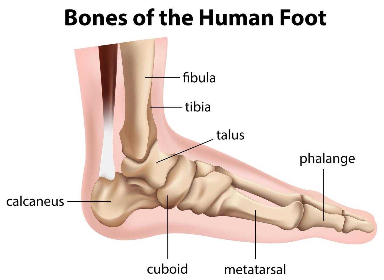
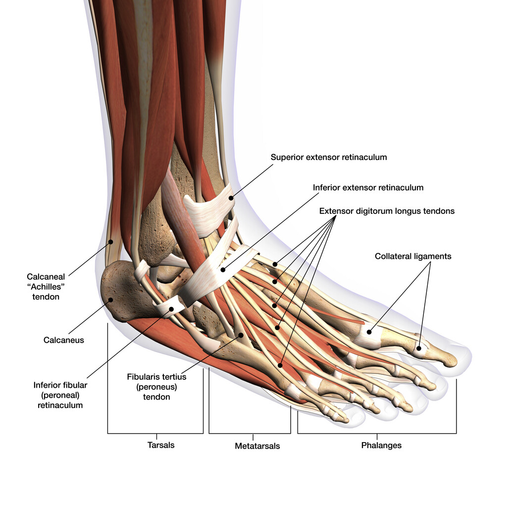
The bones of the foot provide mechanical support for the soft tissues; helping the foot withstand the weight of the body whilst standing and in motion. They can be divided into three groups: Tarsals - a set of seven irregularly shaped bones. They are situated proximally in the foot in the ankle area. Metatarsals - connect the phalanges to.
Bones of the human foot diagram 1142236 Vector Art at Vecteezy
Listed below are 3 common areas of pain in the foot and their causes: Pain in the ball of the foot. Pain in the ball of the foot, located on the bottom of the foot behind the toes, may be caused by nerve or joint damage in that area. In addition, a benign (noncancerous) growth, such as Morton's neuroma, may cause the pain.
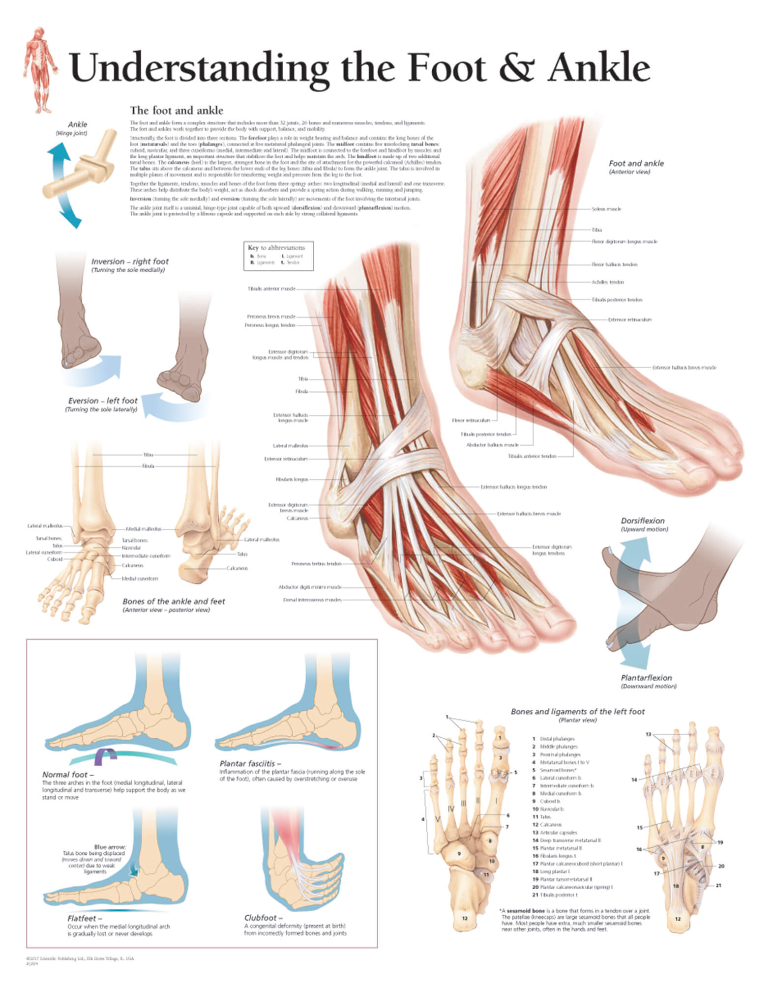
Understanding the Foot & Ankle Scientific Publishing
Figure 1: Bones of the Foot and Ankle Regions of the Foot The foot is traditionally divided into three regions: the hindfoot, the midfoot, and the forefoot (Figure 2). Additionally, the lower leg often refers to the area between the knee and the ankle and this area is critical to the functioning of the foot.

Anatomy Chart Foot and Ankle
The foot pain identifier diagrams you find here will help you to identify the possible causes of your foot problem and then help you find out everything you need to know about the causes, symptoms, diagnosis and best treatment options for each.

Anatomy of the Foot and Ankle OrthoPaedia
Tibia Fibula Talus Cuneiforms Cuboid Navicular Many of the muscles that affect larger foot movements are located in the lower leg. However, the foot itself is a web of muscles that can perform.

Pin on Body (of) Work
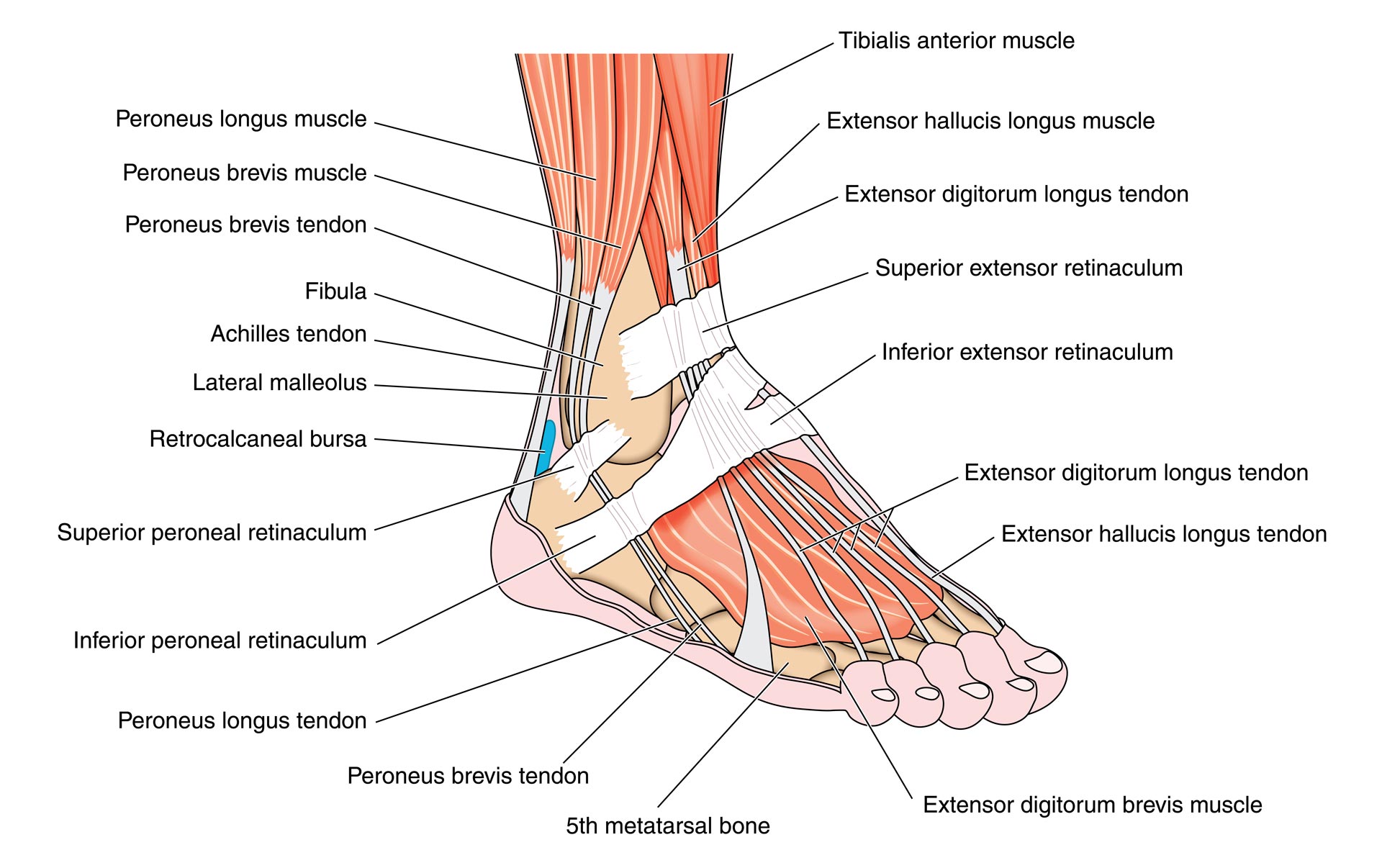
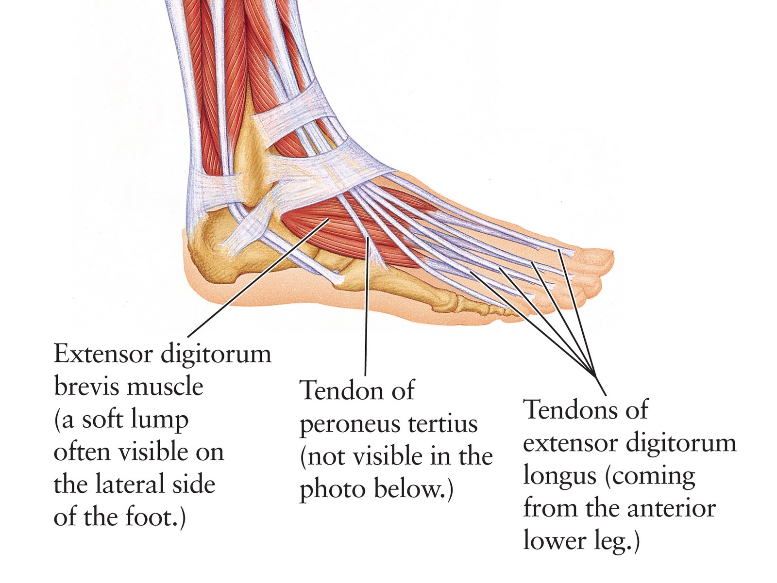
The muscles acting on the foot can be divided into two distinct groups; extrinsic and intrinsic muscles. Extrinsic muscles arise from the anterior, posterior and lateral compartments of the leg. They are mainly responsible for actions such as eversion, inversion, plantarflexion and dorsiflexion of the foot. Intrinsic muscles are located within.

Ankle and Foot Pain Massage Therapy Connections
The foot (pl.: feet) is an anatomical structure found in many vertebrates. It is the terminal portion of a limb which bears weight and allows locomotion. In many animals with feet, the foot is a separate [clarification needed] organ at the terminal part of the leg made up of one or more segments or bones, generally including claws and/or nails.

Foot Also, explore tools to convert foot or centimeter to other
The foot diagram has a complex structure made up of bones, ligaments, muscles, and tendons. Understanding the structure of the foot is best done by looking at a foot diagram where the anatomy has been labeled. If you would like to learn all the parts of the foot structure, you have come to the right place. In this article, we will look at all.
.jpg)
Foot Bone Diagram resource Imageshare
When to see a doctor Summary The foot is an intricate part of the body, consisting of 26 bones, 33 joints, 107 ligaments, and 19 muscles. Scientists group the bones of the foot into the.
Human Anatomy for the Artist The Dorsal Foot How Do I Love Thee? Let
The talus is held in place by the foot bones surrounding it and various ligaments. 4. Calcaneus. The calcaneus is more commonly known as the heel bone. It is the largest of the foot bones and has a quadrangular shape. The calcaneus is the most commonly fractured tarsal bone, usually from a high fall.

Foot Description, Drawings, Bones, & Facts Britannica
The foot can be divided into three regions, the hindfoot, midfoot, and forefoot. F o o t B o n e s L a b e l e d D i a g r a m Names of the Bones in the Foot With Basic Anatomy Tarsal Bones The tarsals are a group of 7 irregular bones forming the hindfoot and the midfoot. These bones are arranged in two rows, proximal and distal.
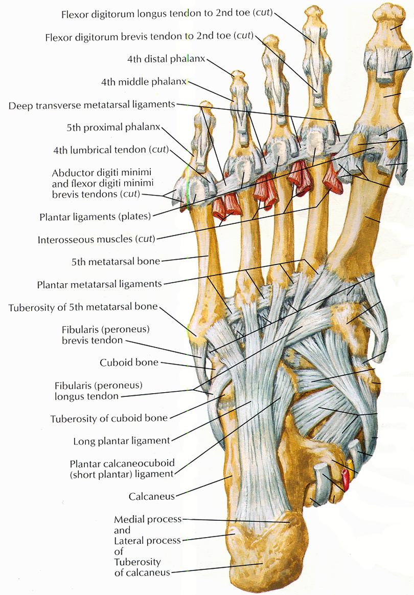
Tendons of the Foot JOI Jacksonville Orthopaedic Institute
Human body Skeletal System Bones of foot Bones of foot The 26 bones of the foot consist of eight distinct types, including the tarsals, metatarsals, phalanges, cuneiforms, talus, navicular,.

Diagrams of the Foot Labeled 101 Diagrams
Morton's neuroma is a common foot problem where compression on a nerve in the ball of the foot causes burning, tingling, and pain near the third and fourth toes. It can make you feel like you have a pebble in your shoe or on a fold in your sock. Wearing high heels is a common cause of Morton's neuroma.
Muscles that lift the Arches of the Feet
Dr. Ebraheim's educational animated video describes anatomical structures of the foot and ankle, The Bony Anatomy, The Joints, Ligaments, and the Compartment.